
Diabetes is the leading cause of blindness in working-age adults in India. The good news: with annual screenings, most vision loss is entirely preventable.
What Is Diabetic Retinopathy?
Diabetic retinopathy is a complication of diabetes that affects the blood vessels in the retina — the light-sensitive tissue at the back of the eye. Chronically elevated blood glucose levels damage these tiny vessels, causing them to swell, leak, or close off entirely. In advanced cases, abnormal new blood vessels grow on the retinal surface, which can bleed and lead to retinal detachment.
It is the most common cause of new blindness among working-age adults in India and globally. India is home to approximately 101 million people with diabetes (the second-highest globally), making diabetic eye disease a major public health priority.
The Four Stages of Diabetic Retinopathy
Diabetic retinopathy progresses through four recognised stages:
The earliest stage. Small areas of balloon-like swelling (microaneurysms) appear in the retinal blood vessels. Vision is usually unaffected at this stage.
More blood vessels that nourish the retina become blocked. Vision may remain normal but the risk of progression is increasing.
Many blood vessels are blocked, depriving several areas of the retina of their blood supply. These areas signal the body to grow new blood vessels.
The most advanced stage. New, fragile blood vessels grow along the retina and vitreous. These can leak blood, causing severe vision loss. Tractional retinal detachment may occur.
Diabetic macular oedema can occur at any stage of diabetic retinopathy. It involves swelling of the macula — the central part of the retina responsible for detailed vision — due to leaking blood vessels. DMO is the most common cause of moderate vision loss in people with diabetes and requires prompt treatment.
Symptoms: Why Diabetic Retinopathy Is So Dangerous
The most dangerous aspect of diabetic retinopathy is that it causes no symptoms in its early stages. Many patients first notice vision changes only when the disease has reached an advanced, harder-to-treat stage. Symptoms that may eventually appear include:
- Spots or dark strings floating in your vision (floaters)
- Blurred or fluctuating vision
- Dark or empty areas in your vision
- Difficulty with colour perception
- Sudden, severe vision loss (in advanced cases with vitreous haemorrhage)
Prevention: Protecting Your Eyes With Diabetes
Research consistently shows that tight control of blood glucose, blood pressure, and blood lipids is the most powerful intervention for preventing diabetic retinopathy or slowing its progression.
Aim for an HbA1c below 7% (or as advised by your physician). Each 1% reduction in HbA1c reduces the risk of microvascular complications by approximately 25–35%.
Target a blood pressure below 130/80 mmHg. Hypertension accelerates retinal blood vessel damage in diabetics.
Elevated lipids contribute to hard exudate deposits in the retina. A healthy diet and appropriate medication help manage this.
Smoking significantly worsens the vascular complications of diabetes, including retinopathy. Cessation at any stage is beneficial.
Regular moderate exercise improves insulin sensitivity and helps maintain healthy blood glucose levels. Aim for at least 150 minutes per week.
This is the single most important action for preserving sight. Annual retinal screening detects changes before vision is affected and enables timely treatment.
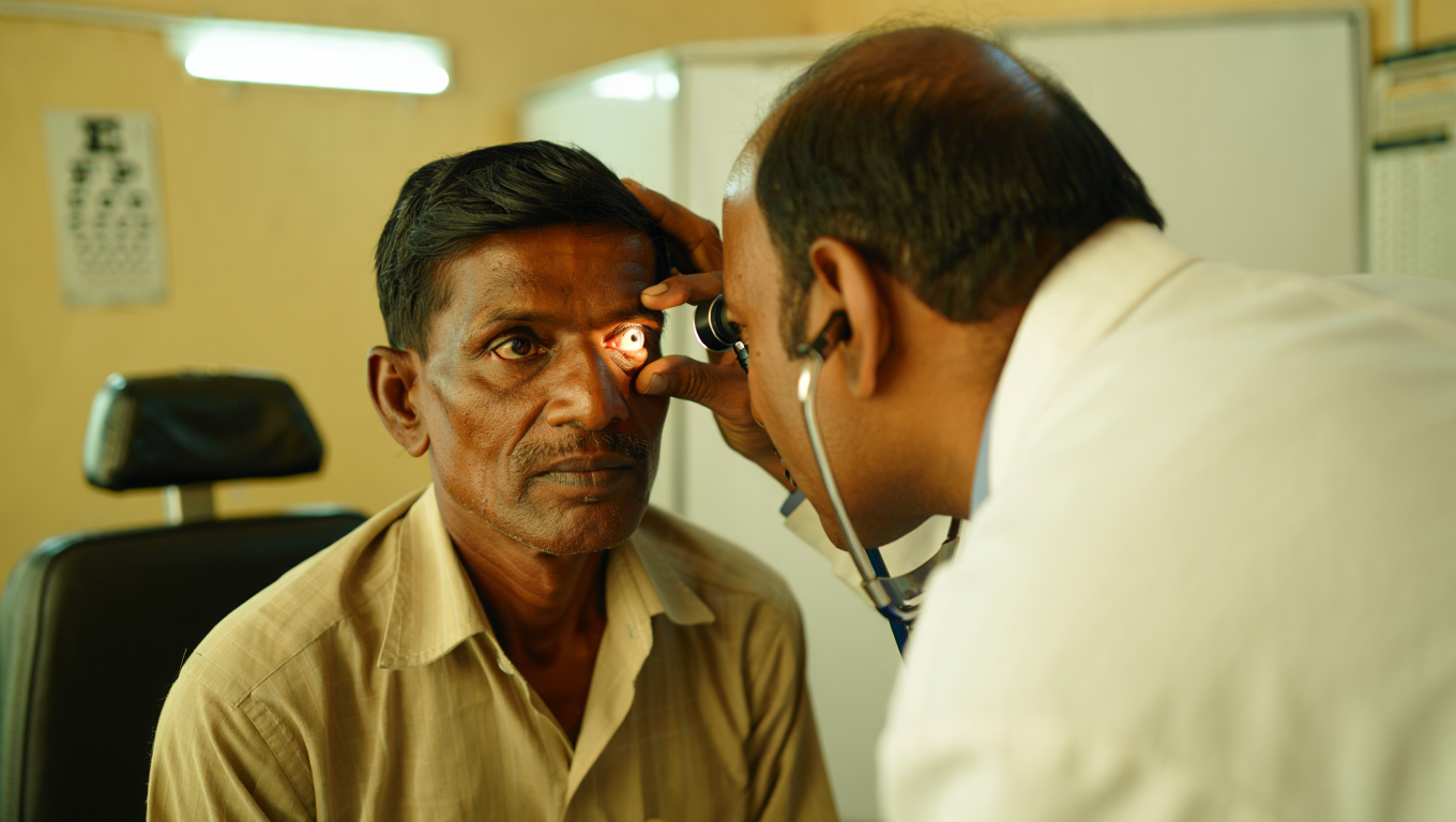
Detection: What Happens at a Diabetic Eye Screening
At Shine Eye Clinic, our diabetic retinal examination includes:
Eye drops widen the pupil for a thorough view of the retina, optic nerve, and blood vessels.
Digital retinal photos provide a baseline for monitoring changes over time.
A cross-sectional scan of the retina that detects macular oedema with extreme precision.
A dye-based test that maps blood vessel leakage and abnormal vessel growth (used in advanced cases).
Treatment Options
Treatment depends on the stage and severity of the disease. Available options include:
Intravitreal injections of anti-VEGF agents (such as ranibizumab or bevacizumab) are now the first-line treatment for diabetic macular oedema and proliferative DR. They block the growth of abnormal blood vessels and reduce leakage. Multiple injections over months to years may be needed.
Laser treatment seals leaking blood vessels and destroys abnormal new vessels. Focal laser is used for DMO; pan-retinal photocoagulation (PRP) is used for proliferative DR. Less commonly used as first-line since anti-VEGF has become available, but remains important in many cases.
Steroid injections or sustained-release implants can reduce macular oedema, particularly in patients who respond inadequately to anti-VEGF. Risk of elevated eye pressure and cataract must be monitored.
Surgical removal of the vitreous gel is necessary for advanced cases involving non-clearing vitreous haemorrhage or tractional retinal detachment. Highly specialised surgery performed by vitreoretinal surgeons.
Who Should Get Screened and When?
First dilated eye exam within 5 years of diagnosis, then annually.
Dilated eye exam at the time of diagnosis (disease may have been present years before detection), then annually.
Eye exam in first trimester; may require more frequent monitoring during pregnancy.
Those with poor glucose control, long disease duration, hypertension, or known retinopathy require more frequent examinations — every 3–6 months in some cases.
The Bottom Line
Diabetic retinopathy is silent, progressive, and largely preventable. If you have diabetes, the most important thing you can do for your eyes is to attend your annual retinal screening — even if your vision feels perfectly fine. Caught early, this condition can be monitored and treated effectively. Left undetected, it can cause irreversible vision loss. Don't wait for symptoms that may never come until it's too late.
Senior Ophthalmologist at Shine Eye Clinic. Specialises in comprehensive eye care, refractive surgery, and paediatric ophthalmology. Committed to making quality eye care accessible to every patient.